We study mechanisms underlying embryonic neurodevelopment and malignancies of prenatal origin.
Our laboratory studies the cellular and molecular mechanisms that control the formation of the nervous system in the embryo. We are interested in the communication of progenitor and neuronal cells with their environment, with a focus on the cellular processes and molecular signaling that control the colonization of embryonic territories by migrating neural cells and axons. We are currently addressing these questions by studying a remarkable embryonic cell population, the neural crest, at the origin of multiple derivatives, including the sympathetic chain, adrenal medulla, and enteric nervous system. Using experimental manipulations in the avian embryo model and transcriptomics, we study the emergence of enteric neural circuits and brain-gut connectivity to characterize the gene programs that mediate communication between cells and their environment. We are investigating whether these programs are conserved in the human embryo and whether their deregulation might contribute to gut neurodevelopmental disorders.
In parallel, we study pediatric cancers in the light of their embryonic origin, in particular neural crest derived neuroblastoma and cerebellar precursor-derived medulloblastoma. The heterogeneity and plasticity that characterize these malignancies and underlie their aggressiveness are thought to be rooted in the embryonic context of their emergence. Tumorigenic events take place in cells that actively communicate with their environment and are endowed with the proliferative and migratory properties necessary for tissue formation.
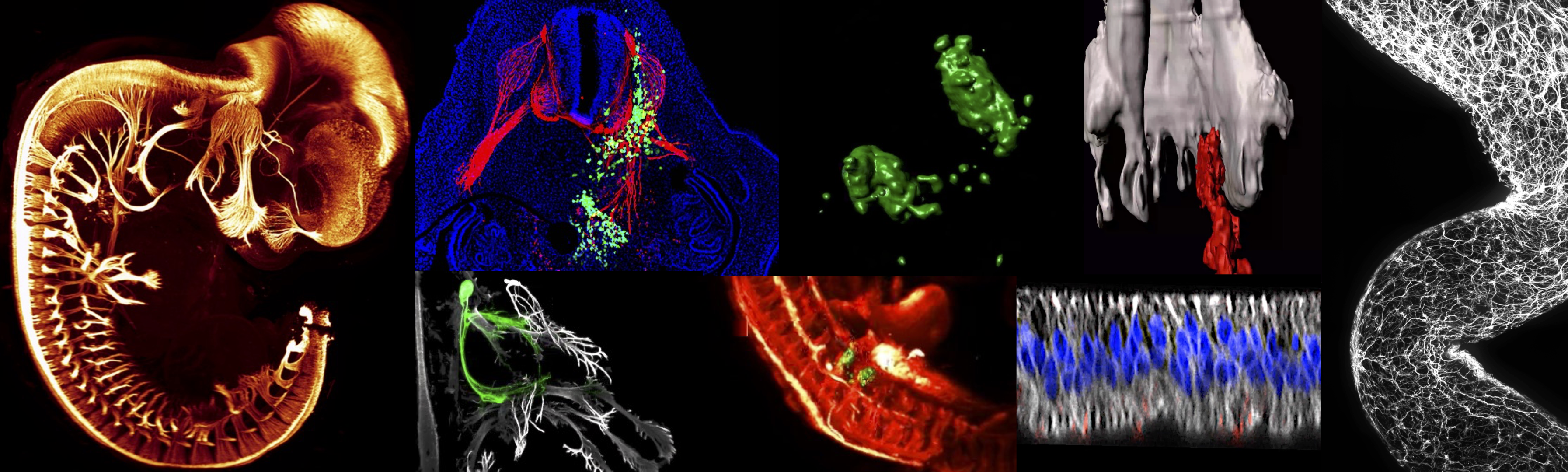
While becoming malignant, tumoral cells retain many of the characteristics of the cells of origin. Our goal is to understand how during tumorigenesis and metastasis this dual physiological and tumorigenic potential is manifested by the opportunistic exploitation or hijacking of developmental mechanisms and signaling pathways. This basic research will pave the way for the development of therapies that specifically target tumor-specific behavior and signaling. To address these questions, and taking advantage of our developmental biology models, we have established a specific in vivo paradigm that recapitulates the embryonic context for malignant cells. It consists of transplanting human tumor cells into selected tissues of the avian embryo. Our studies are based on a multi-approach strategy combining experimental embryology, functional studies of genes of interest in avian models, 3D light sheet microscopy to map cells and molecules at the whole embryo level, videomicroscopy, and large-scale transcriptomic analyses.
For the general public
How are our neural circuits built? the newborn neuron develops an extension, the axon, destined to embark on an incredible journey in search of the cells with which it will establish communication. Thus, during embryonic and postnatal development, millions of axons go in search of their partners, some remaining confined to the brain or spinal cord, others colonizing the whole organism to innervate the muscles, the skin, the viscera. Many neuronal cells also migrate to colonize distal territories where they will form a nervous structure. Molecular signals are expressed in embryonic tissues that allow axons and cells to locate themselves in space. They are called topographic or guidance signals. Getting each axon and cell to its destination is a real challenge, and it is these processes that our team is studying. Can axons and cells get lost or misdirected along the way? Alterations of cell and axon migration are responsible for childhood pathologies. Many remain undiscovered because the identification of alterations of these early processes remains difficult. Cells can also become malignant prenatally, at a time when they are undergoing migration and proliferation. They give rise to tumors that can be highly metastatic and have a poor outcome. Our team studies the behavior of malignant cells and how they communicate with embryonic tissues during tumorigenesis and metastasis.
South-ROCK
Our team is a member of the South-ROCK integrated research center of excellence in pediatric oncology of Lyon-Marseille, one of the three laureates of the French National Cancer Institute's PEDIACRIEX call for projects.
REACT4KIDS
Our team is part of REACT4KIDS (REsearchers in oncology ACTing for kids), a national collaborative network of pediatric oncology research laboratories whose goal is to foster collaborative research for gaining knowledge about the biology of childhood cancers to pave the way for innovative therapies tailored to these cancers.
Start Up
Oncofactory is a start-up founded in 2016 as a spin-off of the Castellani lab, based on the work of Dr. Céline Delloye Bourgeois and Dr. Valérie Castellani at the NeuroMyoGene Institute in Lyon. Oncofactory offers an innovative in vivo platform suited for all cancers consisting in the creation of miniaturized replicas of patient tumors in an embryonic organism. Tumors are analyzed in the whole organism using 3D microscopy. Replicas can be collected back for a range of large scale molecular analysis. Using its unique technological platform Oncofactory evaluates the efficacy, studies the mechanisms of action of therapeutic candidates, helps the design of combi-therapies through a dedicated program combining transcriptomic analysis and its model of patient tumors replicas.
Céline Delloye-Bourgeois — Group leader, Center of Research of Cancerology of Lyon, France
Homaira Nawabi — Group leader, Grenoble Institute of Neuroscience, France
Camille Charoy — Senior microscopist at The Francis Crick Institute, UK
Florie Reynaud — Research Engineer, Center of Research of Cancerology of Lyon, France
Elise Arbeille — Assistant professor, University of Méditerranée, Marseille, France
Anne Briançon-Marjollet — Assistant professor, Grenoble-Alpes University, Grenoble, France
Selected publications
Functional precision oncology for follicular lymphoma with patient-derived xenograft in avian embryos. Zala M, Lipinski B, Costechareyre C, Jarrosson L, Teinturier R, Julia E, Lacourrège M, Verney A, Guitton J, Traverse-Glehen A, Bachy E, Salles G, Huet S, Genestier L, Castellani V, Delloye-Bourgeois C, Sujobert P. Leukemia (2024) — Show abstract
An in vivo avian model of human melanoma to perform rapid and robust preclinical studies Jarrosson L, Dalle S, Costechareyre C, Tang Y, Grimont M, Plaschka M, Lacourrège M, Teinturier R, Le Bouar M, Maucort-Boulch D, Eberhardt A, Castellani V, Caramel J, Delloye-Bourgeois C. EMBO Mol Med. (2023) — Show abstract
Metastatic melanoma patients carrying a BRAFV600 mutation can be treated with a combination of BRAF and MEK inhibitors (BRAFi/MEKi), but innate and acquired resistance invariably occurs. Predicting patient response to targeted therapies is crucial to guide clinical decision. We describe here the development of a highly efficient patient-derived xenograft model adapted to patient melanoma biopsies, using the avian embryo as a host (AVI-PDX). In this in vivo paradigm, we depict a fast and reproducible tumor engraftment of patient samples within the embryonic skin, preserving key molecular and phenotypic features. We show that sensitivity and resistance to BRAFi/MEKi can be reliably modeled in these AVI-PDX , as well as synergies with other drugs. We further provide proof-of-concept that the AVI-PDX models the diversity of responses of melanoma patients to BRAFi/MEKi, within days, hence positioning it as a valuable tool for the design of personalized medicine assays and for the evaluation of novel combination strategies.
A balance of noncanonical Semaphorin signaling from the cerebrospinal fluid regulates apical cell dynamics during corticogenesis. Gerstmann K, Kindbeiter K, Telley L, Bozon M, Reynaud F, Théoulle E, Charoy C, Jabaudon D, Moret F, Castellani V. Sci Advances (2022) — Show abstract
During corticogenesis, dynamic regulation of apical adhesion is fundamental to generate correct numbers and cell identities. While radial glial cells (RGCs) maintain basal and apical anchors, basal progenitors and neurons detach and settle at distal positions from the apical border. Whether diffusible signals delivered from the cerebrospinal fluid (CSF) contribute to the regulation of apical adhesion dynamics remains fully unknown. Secreted class 3 Semaphorins (Semas) trigger cell responses via Plexin-Neuropilin (Nrp) membrane receptor complexes. Here, we report that unconventional Sema3-Nrp preformed complexes are delivered by the CSF from sources including the choroid plexus to Plexin-expressing RGCs via their apical endfeet. Through analysis of mutant mouse models and various ex vivo assays mimicking ventricular delivery to RGCs, we found that two different complexes, Sema3B/Nrp2 and Sema3F/Nrp1, exert dual effects on apical endfeet dynamics, nuclei positioning, and RGC progeny. This reveals unexpected balance of CSF-delivered guidance molecules during cortical development.
GPC3-Unc5 receptor complex structure and role in cell migration Akkermans O, Delloye-Bourgeois C, Peregrina C, Carrasquero-Ordaz M, Kokolaki M, Berbeira-Santana M, Chavent M, Reynaud F, Raj R, Agirre J, Aksu M, White ES, Lowe E, Ben Amar D, Zaballa S, Huo J, Pakos I, McCubbin PTN, Comoletti D, Owens RJ, Robinson CV, Castellani V, Del Toro D, Seiradake E. Cell (2022) — Show abstract
Neural migration is a critical step during brain development that requires the interactions of cell-surface guidance receptors. Cancer cells often hijack these mechanisms to disseminate. Here, we reveal crystal structures of Uncoordinated-5 receptor D (Unc5D) in complex with morphogen receptor glypican-3 (GPC3), forming an octameric glycoprotein complex. In the complex, four Unc5D molecules pack into an antiparallel bundle, flanked by four GPC3 molecules. Central glycan-glycan interactions are formed by N-linked glycans emanating from GPC3 (N241 in human) and C-mannosylated tryptophans of the Unc5D thrombospondin-like domains. MD simulations, mass spectrometry and structure-based mutants validate the crystallographic data. Anti-GPC3 nanobodies enhance or weaken Unc5-GPC3 binding and, together with mutant proteins, show that Unc5/GPC3 guide migrating pyramidal neurons in the mouse cortex, and cancer cells in an embryonic xenograft neuroblastoma model. The results demonstrate a conserved structural mechanism of cell guidance, where finely balanced Unc5-GPC3 interactions regulate cell migration.
Environmental cues from neural crest derivatives act as metastatic triggers in an embryonic neuroblastoma model. Ben Amar D, Thoinet K, Villalard B, Imbaud O, Costechareyre C, Jarrosson L, Reynaud F, Novion Ducassou J, Couté Y, Brunet JF, Combaret V, Corradini N, Delloye-Bourgeois C, Castellani V. Nature Communications (2022) — Show abstract
Embryonic malignant transformation is concomitant to organogenesis, often affecting multipotent and migratory progenitors. While lineage relationships between malignant cells and their physiological counterparts are extensively investigated, the contribution of exogenous embryonic signals is not fully known. Neuroblastoma (NB) is a childhood malignancy of the peripheral nervous system arising from the embryonic trunk neural crest (NC) and characterized by heterogeneous and interconvertible tumor cell identities. Here, using experimental models mimicking the embryonic context coupled to proteomic and transcriptomic analyses, we show that signals released by embryonic sympathetic ganglia, including Olfactomedin-1, induce NB cells to shift from a noradrenergic to mesenchymal identity, and to activate a gene program promoting NB metastatic onset and dissemination. From this gene program, we extract a core signature specifically shared by metastatic cancers with NC origin. This reveals non-cell autonomous embryonic contributions regulating the plasticity of NB identities and setting pro-dissemination gene programs common to NC-derived cancers.
An avian embryo patient-derived xenograft model for preclinical studies of human breast cancers. Jarrosson L, Costechareyre C, Gallix F, Ciré S, Gay F, Imbaud O, Teinturier R, Marangoni E, Aguéra K, Delloye-Bourgeois C, Castellani V. iScience (2021) — Show abstract
Lack of preclinical patient-derived xenograft cancer models in which to conduct large-scale molecular studies seriously impairs the development of effective personalized therapies. We report here an in vivo concept consisting of implanting human tumor cells in targeted tissues of an avian embryo, delivering therapeutics, evaluating their efficacy by measuring tumors using light sheet confocal microscopy, and conducting large-scale RNA-seq analysis to characterize therapeutic-induced changes in gene expression. The model was established to recapitulate triple-negative breast cancer (TNBC) and validated using TNBC standards of care and an investigational therapeutic agent.
X-linked partial corpus callosum agenesis with mild intellectual disability: identification of a novel L1CAM pathogenic variant. Bousquet I, Bozon M, Castellani V, Touraine R, Piton A, Gérard B, Guibaud L, Sanlaville D, Edery P, Saugier-Veber P, Putoux A. Neurogenetics (2021) — Show abstract
athogenic variants in L1CAM, the gene encoding the L1 cell adhesion molecule, are responsible for a wide clinical spectrum including X-linked hydrocephalus with stenosis of the Sylvius aqueduct, MASA syndrome (mental retardation, aphasia, shuffling gait, adducted thumbs), and a form of spastic paraplegia (SPG1). A moderate phenotype with mild intellectual disability (ID) and X-linked partial corpus callosum agenesis (CCA) has only been related to L1CAM in one family. We report here a second family, including 5 patients with mild to moderate ID and partial CCA without signs usually associated with L1CAM pathogenic variations (such as hydrocephalus, pyramidal syndrome, thumb adductus, aphasia). We identified a previously unreported c.3226A > C transversion leading to a p.Thr1076Pro amino acid substitution in the fifth fibronectin type III domain (FnIII) of the protein which co-segregates with the phenotype within the family. We performed in vitro assays to assess the pathogenic status of this variation. First, the expression of the novel p.Thr1076Pro mutant in COS7 cells resulted in endoplasmic reticulum (ER) retention and reduced L1CAM cell surface expression, which is expected to affect both L1CAM-mediated cell-cell adhesion and neurite growth. Second, immunoblotting techniques showed that the immature form of the L1CAM protein was increased, indicating that this variation led to a lack of maturation of the protein. ID associated with CCA is not a common clinical presentation of L1CAM pathogenic variants. Genome-wide analyses will identify such variations and it is important to acknowledge this atypical phenotype.
SlitC-PlexinA1 mediates iterative inhibition for orderly passage of spinal commissural axons through the floor plate. Ducuing H, Gardette T, Pignata A, Kindbeiter K, Bozon M, Thoumine O, Delloye-Bourgeois C, Tauszig-Delamasure S, Castellani V. Elife (2020) — Show abstract
Spinal commissural axon navigation across the midline in the floor plate requires repulsive forces from local Slit repellents. The long-held view is that Slits push growth cones forward and prevent them from turning back once they became sensitized to these cues after midline crossing. We analyzed with fluorescent reporters Slits distribution and FP glia morphology. We observed clusters of Slit-N and Slit-C fragments decorating a complex architecture of glial basal process ramifications. We found that PC2 proprotein convertase activity contributes to this pattern of ligands. Next, we studied Slit-C acting via PlexinA1 receptor shared with another FP repellent, the Semaphorin3B, through generation of a mouse model baring PlexinA1Y1815F mutation abrogating SlitC but not Sema3B responsiveness, manipulations in the chicken embryo, and ex vivo live imaging. This revealed a guidance mechanism by which SlitC constantly limits growth cone exploration, imposing ordered and forward-directed progression through aligned corridors formed by FP basal ramifications.
A Spatiotemporal Sequence of Sensitization to Slits and Semaphorins Orchestrates Commissural Axon Navigation. Pignata A, Ducuing H, Boubakar L, Gardette T, Kindbeiter K, Bozon M, Tauszig-Delamasure S, Falk J, Thoumine O, Castellani V. Cell Reports (2019) — Show abstract
Accurate perception of guidance cues is crucial for cell and axon migration. During initial navigation in the spinal cord, commissural axons are kept insensitive to midline repellents. Upon midline crossing in the floor plate, they switch on responsiveness to Slit and Semaphorin repulsive signals and are thus propelled away and prevented from crossing back. Whether and how the different midline repellents control specific aspects of this navigation remain to be elucidated. We set up a paradigm for live-imaging and super-resolution analysis of PlexinA1, Neuropilin-2, and Robo1/2 receptor dynamics during commissural growth cone navigation in chick and mouse embryos. We uncovered a remarkable program of sensitization to midline cues achieved by unique spatiotemporal sequences of receptor allocation at the growth-cone surface that orchestrates receptor-specific growth-cone behavior changes. This reveals post-translational mechanisms whereby coincident guidance signals are temporally resolved to allow the generation of specific guidance responses.
Hijacking of Embryonic Programs by Neural Crest-Derived Neuroblastoma: From Physiological Migration to Metastatic Dissemination. Delloye-Bourgeois C, Castellani V. Front Mol Neurosci. (2019) — Show abstract
In the developing organism, complex molecular programs orchestrate the generation of cells in adequate numbers, drive them to migrate along the correct pathways towards appropriate territories, eliminate superfluous cells, and induce terminal differentiation of survivors into the appropriate cell-types. Despite strict controls constraining developmental processes, malignancies can emerge in still immature organisms. This is the case of neuroblastoma (NB), a highly heterogeneous disease, predominantly affecting children before the age of 5 years. Highly metastatic forms represent half of the cases and are diagnosed when disseminated foci are detectable. NB arise from a transient population of embryonic cells, the neural crest (NC), and especially NC committed to the establishment of the sympatho-adrenal tissues. The NC is generated at the dorsal edge of the neural tube (NT) of the vertebrate embryo, under the action of NC specifier gene programs. NC cells (NCCs) undergo an epithelial to mesenchymal transition, and engage on a remarkable journey in the developing embryo, contributing to a plethora of cell-types and tissues. Various NCC sub-populations and derived lineages adopt specific migratory behaviors, moving individually as well as collectively, exploiting the different embryonic substrates they encounter along their path. Here we discuss how the specific features of NCC in development are re-iterated during NB metastatic behaviors.
Septin functions during neuro-development, a yeast perspective Falk J, Boubakar L, Castellani V. Curr Opin Neurobiol (2019) — Show abstract
Septins, discovered almost half a century ago in yeast, have prominent contributions in a broad range of morphological and functional processes from yeast to human. Septins now emerge as key players of neurodevelopment and more specifically of the mechanisms driving the complex morphological differentiation and compartmentalization of neurons that are fundamental to their function. We review here recent advances in Septin-mediated processes of neuron differentiation, which enlighten similarities and differences between neuron and yeast polarity programs.
Commissural axon navigation in the spinal cord: A repertoire of repulsive forces is in command. Ducuing H, Gardette T, Pignata A, Tauszig-Delamasure S, Castellani V. Semin Cell Dev Biol. (2019) — Show abstract
The navigation of commissural axons in the developing spinal cord has attracted multiple studies over the years. Many important concepts emerged from these studies which have enlighten the general mechanisms of axon guidance. The navigation of commissural axons is regulated by a series of cellular territories which provides the diverse guidance information necessary to ensure the successive steps of their pathfinding towards, across, and away from the ventral midline. In this review, we discuss how repulsive forces, by propelling, channelling, and confining commissural axon navigation, bring key contributions to the formation of this neuronal projection.
Microenvironment-Driven Shift of Cohesion/Detachment Balance within Tumors Induces a Switch toward Metastasis in Neuroblastoma. Delloye-Bourgeois C, Bertin L, Thoinet K, Jarrosson L, Kindbeiter K, Buffet T, Tauszig-Delamasure S, Bozon M, Marabelle A, Combaret V, Bergeron C, Derrington E, Castellani V. Cancer Cell (2017) — Show abstract
Neuroblastoma (NB) is a childhood cancer arising from sympatho-adrenal neural crest cells. Disseminated forms have high frequency of multiple tumoral foci whose etiology remains unknown; NB embryonic origin limits investigations in patients and current models. We developed an avian embryonic model driving human NB tumorigenesis in tissues homologous to patients. We found that aggressive NBs display a metastatic mode, secondary dissemination via peripheral nerves and aorta. Through tumor transcriptional profiling, we found that NB dissemination is induced by the shutdown of a pro-cohesion autocrine signal, SEMA3C, which constrains the tumoral mass. Lowering SEMA3C levels shifts the balance toward detachment, triggering NB cells to collectively evade the tumor. Together with patient cohort analysis, this identifies a microenvironment-driven pro-metastatic switch for NB.
Molecular Memory of Morphologies by Septins during Neuron Generation Allows Early Polarity Inheritance. Boubakar L, Falk J, Ducuing H, Thoinet K, Reynaud F, Derrington E, Castellani V. Neuron (2017) — Show abstract
Transmission of polarity established early during cell lineage history is emerging as a key process guiding cell differentiation. Highly polarized neurons provide a fascinating model to study inheritance of polarity over cell generations and across morphological transitions. Neural crest cells (NCCs) migrate to the dorsal root ganglia to generate neurons directly or after cell divisions in situ. Using live imaging of vertebrate embryo slices, we found that bipolar NCC progenitors lose their polarity, retracting their processes to round for division, but generate neurons with bipolar morphology by emitting processes from the same locations as the progenitor. Monitoring the dynamics of Septins, which play key roles in yeast polarity, indicates that Septin 7 tags process sites for re-initiation of process growth following mitosis. Interfering with Septins blocks this mechanism. Thus, Septins store polarity features during mitotic rounding so that daughters can reconstitute the initial progenitor polarity.
Genetic specification of left-right asymmetry in the diaphragm muscles and their motor innervation. Charoy C, Dinvaut S, Chaix Y, Morlé L, Sanyas I, Bozon M, Kindbeiter K, Durand B, Skidmore JM, De Groef L, Seki M, Moons L, Ruhrberg C, Martin JF, Martin DM, Falk J, Castellani V. Elife (2017) — Show abstract
The diaphragm muscle is essential for breathing in mammals. Its asymmetric elevation during contraction correlates with morphological features suggestive of inherent left-right (L/R) asymmetry. Whether this asymmetry is due to L versus R differences in the muscle or in the phrenic nerve activity is unknown. Here, we have combined the analysis of genetically modified mouse models with transcriptomic analysis to show that both the diaphragm muscle and phrenic nerves have asymmetries, which can be established independently of each other during early embryogenesis in pathway instructed by Nodal, a morphogen that also conveys asymmetry in other organs. We further found that phrenic motoneurons receive an early L/R genetic imprint, with L versus R differences both in Slit/Robo signaling and MMP2 activity and in the contribution of both pathways to establish phrenic nerve asymmetry. Our study therefore demonstrates L-R imprinting of spinal motoneurons and describes how L/R modulation of axon guidance signaling helps to match neural circuit formation to organ asymmetry.
Cerebrospinal fluid-derived Semaphorin3B orients neuroepithelial cell divisions in the apicobasal axis. Arbeille E, Reynaud F, Sanyas I, Bozon M, Kindbeiter K, Causeret F, Pierani A, Falk J, Moret F, Castellani V. Nature Communications (2015) — Show abstract
The spatial orientation of cell divisions is fundamental for tissue architecture and homeostasis. Here we analysed neuroepithelial progenitors in the developing mouse spinal cord to determine whether extracellular signals orient the mitotic spindle. We report that Semaphorin3B (Sema3B) released from the floor plate and the nascent choroid plexus in the cerebrospinal fluid (CSF) controls progenitor division orientation. Delivery of exogenous Sema3B to neural progenitors after neural tube opening in living embryos promotes planar orientation of their division. Preventing progenitor access to cues present in the CSF by genetically engineered canal obstruction affects the proportion of planar and oblique divisions. Sema3B knockout phenocopies the loss of progenitor access to the CSF. Sema3B binds to the apical surface of mitotic progenitors and exerts its effect via Neuropilin receptors, GSK3 activation and subsequent inhibition of the microtubule stabilizer CRMP2. Thus, extrinsic control mediated by the Semaphorin signalling orients progenitor divisions in neurogenic zones.
PlexinA1 is a new Slit receptor and mediates axon guidance function of Slit C-terminal fragments. Delloye-Bourgeois C, Jacquier A, Charoy C, Reynaud F, Nawabi H, Thoinet K, Kindbeiter K, Yoshida Y, Zagar Y, Kong Y, Jones YE, Falk J, Chédotal A, Castellani V. Nature Neuroscience (2014) — Show abstract
Robo-Slit and Plexin-Semaphorin signaling participate in various developmental and pathogenic processes. During commissural axon guidance in the spinal cord, chemorepulsion by Semaphorin3B and Slits controls midline crossing. Slit processing generates an N-terminal fragment (SlitN) that binds to Robo1 and Robo2 receptors and mediates Slit repulsive activity, as well as a C-terminal fragment (SlitC) with an unknown receptor and bioactivity. We identified PlexinA1 as a Slit receptor and found that it binds the C-terminal Slit fragment specifically and transduces a SlitC signal independently of the Robos and the Neuropilins. PlexinA1-SlitC complexes are detected in spinal cord extracts, and ex vivo, SlitC binding to PlexinA1 elicits a repulsive commissural response. Analysis of various ligand and receptor knockout mice shows that PlexinA1-Slit and Robo-Slit signaling have complementary roles during commissural axon guidance. Thus, PlexinA1 mediates both Semaphorin and Slit signaling, and Slit processing generates two active fragments, each exerting distinct effects through specific receptors.