Our work focuses on understanding how skeletal muscles are formed and repaired in vertebrates.
We are using chick and mouse to address two main lines of investigation. • Understanding how individual stem cells engage into differentiation or remain in a non-differentiated, quiescent and/or self-renewing state. • To characterize the gene networks underlying the fusion of myoblasts into muscle fibers during embryonic development and muscle repair. This in turn will allow developing the tools and concepts to utilize cell fusion as a mean to repair ailing muscles in heritable muscle diseases (myopathies).
In recent years, our laboratory has focused much effort on understanding the molecular and cellular mechanisms regulating muscle cell fusion. The fusion of differentiating muscle cells to existing muscle fibers is a crucial step of muscle formation and repair that is poorly understood. We have undertaken a genome-wide functional screen on a mouse muscle cell line and identified hundreds of molecules implicated in the fusion of this cell line, with no effect on their proliferation or differentiation. Inhibitors and activators of fusion, members of various signaling pathways, genes that when mutated, lead to muscle dystrophies in human: there are many surprises within this list of putative modulators of muscle fusion. To test their function during fusion, we use the chick embryo as a model. The amenability of the chick embryo to manipulation and imaging, combined with the powerful technique of in vivo electroporation and the strong similarities of muscle formation in birds and mammals provide a unique paradigm to characterize this process in amniotes.
A second line of research is to use skeletal muscle formation in the chick embryo as a model to understand how cells within tissues display complex behaviours while being exposed to an ever-changing cellular environment. We have recently shown that in avian embryos, muscle formation is initiated by Delta1-positive neural crest cells migrating from the dorsal neural tube that, in passing, trigger NOTCH signalling and myogenesis in selected epithelial somite progenitor cells, allowing them to migrate into the nascent muscle to differentiate.
Preliminary data we have now obtained further indicate that in somite cells, the activation of the NOTCH pathway triggers a “signalling module” that couples the initiation of myogenesis with the epithelial-mesenchymal transition (EMT) that allows them to migrate into the growing muscle. This is a significant discovery: in many cellular contexts, essential cell fate decisions are associated with an EMT. This is true at many stages of embryonic development (e.g. the formation of the three germ layers during gastrulation, the formation of neural crest, etc.), but also during pathologies like the metastatic progression of carcinomas. Inhibiting EMT arrests cell fate decision in these experimental models, suggesting a mechanistic link between both processes that has never been understood. Our working hypothesis is that the signalling module we have uncovered underlies the coupling cell fate changes to EMT in a variety of developmental and pathological processes.
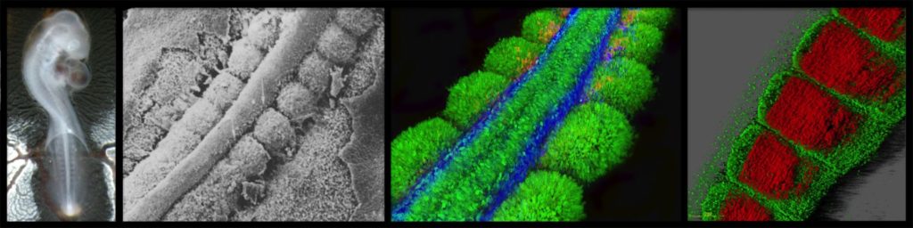
Chicken embryo at 5.5 days of development, clarified by the “3DISCO” technique, observed with a light sheet microscope (Z1 Zeiss, CIQLE). Green: neural crest and peripheral nervous system (anti-HNK1); Blue: dermomyotome, muscle progenitors and dorsal neural tube (anti-PAX7); Red: differentiated muscles (anti-Myosin Heavy Chain). Marie-Julie Dejardin & Christophe Marcelle.
This animation movie shows the morphogenesis and growth of the early myotome (i.e. the primitive muscle) in a chicken embryo. All muscles of the body and limbs derive from somites, which are epithelial balls of cells that form sequentially on both sides of the neural tube as the embryo develops. Shown here is the dorsal compartment of somites, named the dermomyotome, from which trunk muscles derive. In a first stage, cells from the medial, the posterior, the anterior and finally the lateral borders of the somite translocate below the dermomyotome, where they elongate parallel to the antero-posterior axis of the embryo. These elongated, mono-nucleated, post-mitotic cells are called myocytes and together they form what we have named the primary myotome. In a second stage, the central portion of the epithelial dermomyotome undergoes and epithelial-mensenchymal transition (EMT). As a result, part of the dermomyotomal cells can migrate towards the ectoderm to later form the dermis, while other cells are “parachuted” into the primary myotome. Unlike myocytes that do not divide, the parachuted cells are true muscle progenitors, and they can either differentiate or self-renew. Through this process, the muscles can grow during embryonic and fetal life. The muscle stem cells of the adult (named satellite cells) derive from the same population of progenitors identified here. It is important to realize that the same morphogenetic process takes places in mice, and therefore presumably in human. This animation movie was created in 2005 by Jérôme Gros with the free open source 3D software Blender. Publications associated with this movie: Gros, Scaal & Marcelle, Developmental Cell, 2004. Gros, Manceau, Thomé & Marcelle, Nature, 2005. Gros, Serralbo & Marcelle, Nature, 2009.
Transgenic quails reveal dynamic TCF/β-catenin signaling during avian embryonic development. Barzilai-Tutsch H, Morin V, Toulouse G, Chernyavskiy O, Firth S, Marcelle C, Serralbo O. Elife (2022) — Show abstract
The Wnt/β-catenin signaling pathway is highly conserved throughout evolution, playing crucial roles in several developmental and pathological processes. Wnt ligands can act at a considerable distance from their sources and it is therefore necessary to examine not only the Wnt-producing but also the Wnt-receiving cells and tissues to fully appreciate the many functions of this pathway. To monitor Wnt activity, multiple tools have been designed which consist of multimerized Wnt signaling response elements (TCF/LEF binding sites) driving the expression of fluorescent reporter proteins (e.g. GFP, RFP) or of LacZ. The high stability of those reporters leads to a considerable accumulation in cells activating the pathway, thereby making them easily detectable. However, this makes them unsuitable to follow temporal changes of the pathway's activity during dynamic biological events. Even though fluorescent transcriptional reporters can be destabilized to shorten their half-lives, this dramatically reduces signal intensities, particularly when applied in vivo. To alleviate these issues, we developed two transgenic quail lines in which high copy number (12× or 16×) of the TCF/LEF binding sites drive the expression of destabilized GFP variants. Translational enhancer sequences derived from viral mRNAs were used to increase signal intensity and specificity. This resulted in transgenic lines efficient for the characterization of TCF/β-catenin transcriptional dynamic activities during embryogenesis, including using in vivo imaging. Our analyses demonstrate the use of this transcriptional reporter to unveil novel aspects of Wnt signaling, thus opening new routes of investigation into the role of this pathway during amniote embryonic development.
TGFβ signalling acts as a molecular brake of myoblast fusion. Melendez J, Sieiro D, Salgado D, Morin V, Dejardin MJ, Zhou C, Mullen AC, Marcelle C. Nature Communications (2021) — Show abstract
Fusion of nascent myoblasts to pre-existing myofibres is critical for skeletal muscle growth and repair. The vast majority of molecules known to regulate myoblast fusion are necessary in this process. Here, we uncover, through high-throughput in vitro assays and in vivo studies in the chicken embryo, that TGFβ (SMAD2/3-dependent) signalling acts specifically and uniquely as a molecular brake on muscle fusion. While constitutive activation of the pathway arrests fusion, its inhibition leads to a striking over-fusion phenotype. This dynamic control of TGFβ signalling in the embryonic muscle relies on a receptor complementation mechanism, prompted by the merging of myoblasts with myofibres, each carrying one component of the heterodimer receptor complex. The competence of myofibres to fuse is likely restored through endocytic degradation of activated receptors. Altogether, this study shows that muscle fusion relies on TGFβ signalling to regulate its pace.
Transgenesis and web resources in quail. Serralbo O, Salgado D, Véron N, Cooper C, Dejardin MJ, Doran T, Gros J, Marcelle C. Elife (2020) — Show abstract
Due to its amenability to manipulations, to live observation and its striking similarities to mammals, the chicken embryo has been one of the major animal models in biomedical research. Although it is technically possible to genome-edit the chicken, its long generation time (6 months to sexual maturity) makes it an impractical lab model and has prevented it widespread use in research. The Japanese quail (Coturnix coturnix japonica) is an attractive alternative, very similar to the chicken, but with the decisive asset of a much shorter generation time (1.5 months). In recent years, transgenic quail lines have been described. Most of them were generated using replication-deficient lentiviruses, a technique that presents diverse limitations. Here, we introduce a novel technology to perform transgenesis in quail, based on the in vivo transfection of plasmids in circulating Primordial Germ Cells (PGCs). This technique is simple, efficient and allows using the infinite variety of genome engineering approaches developed in other models. Furthermore, we present a website centralizing quail genomic and technological information to facilitate the design of genome-editing strategies, showcase the past and future transgenic quail lines and foster collaborative work within the avian community.
Cytoplasmic NOTCH and membrane derived β-catenin link fate choice to epithelial-mesenchymal transition during myogenesis. Sieiro D, Rios AC, Hirst CE, Marcelle C. Elife (2016) — Show abstract
How cells in the embryo coordinate epithelial plasticity with cell fate decision in a fast changing cellular environment is largely unknown. In chick embryos, skeletal muscle formation is initiated by migrating Delta1-expressing neural crest cells that trigger NOTCH signaling and myogenesis in selected epithelial somite progenitor cells, which rapidly translocate into the nascent muscle to differentiate. Here, we uncovered at the heart of this response a signaling module encompassing NOTCH, GSK-3β, SNAI1 and β-catenin. Independent of its transcriptional function, NOTCH profoundly inhibits GSK-3β activity. As a result SNAI1 is stabilized, triggering an epithelial to mesenchymal transition. This allows the recruitment of β-catenin from the membrane, which acts as a transcriptional co-factor to activate myogenesis, independently of WNT ligand. Our results intimately associate the initiation of myogenesis to a change in cell adhesion and may reveal a general principle for coupling cell fate changes to EMT in many developmental and pathological processes.
A dynamic analysis of muscle fusion in the chick embryo. Sieiro-Mosti D, De La Celle M, Pele M, Marcelle C. Development (2014) — Show abstract
Skeletal muscle development, growth and regeneration depend upon the ability of muscle cells to fuse into multinucleated fibers. Surprisingly little is known about the cellular events that underlie fusion during amniote development. Here, we have developed novel molecular tools to characterize muscle cell fusion during chick embryo development. We show that all cell populations arising from somites fuse, but each with unique characteristics. Fusion in the trunk is slow and independent of fiber length. By contrast, the addition of nuclei in limb muscles is three times more rapid than in trunk and is tightly associated with fiber growth. A complex interaction takes place in the trunk, where primary myotome cells from the medial somite border rarely fuse to one another, but readily do so with anterior and posterior border cells. Conversely, resident muscle progenitors actively fuse with one another, but poorly with the primary myotome. In summary, this study unveils an unexpected variety of fusion behaviors in distinct embryonic domains that is likely to reflect a tight molecular control of muscle fusion in vertebrates.
Migrating cells mediate long-range WNT signaling. Serralbo O, Marcelle C. Development (2014) — Show abstract
In amniotes, it is widely accepted that WNTs secreted by the dorsal neural tube form a concentration gradient that regulates early somite patterning and myotome organization. Here we demonstrate in the chicken embryo that WNT protein is not secreted to act at a distance, but rather loaded onto migrating neural crest cells that deliver it to somites. Inhibiting neural crest migration or ablating their population has a profound impact on the WNT response in somites. Furthermore, we show that a central player in the efficient delivery of WNT to somites is the heparan sulfate proteoglycan GPC4, expressed by neural crest. Together, our data describe a novel mode of signaling whereby WNT proteins hitch a ride on migratory neural crest cells to pattern the somites at a distance from its source.
Neural crest regulates myogenesis through the transient activation of Notch. Rios AC, Serralbo O, Salgado D, Marcelle C. Nature (2011) — Show abstract
How dynamic signalling and extensive tissue rearrangements interact to generate complex patterns and shapes during embryogenesis is poorly understood. Here we characterize the signalling events taking place during early morphogenesis of chick skeletal muscles. We show that muscle progenitors present in somites require the transient activation of NOTCH signalling to undergo terminal differentiation. The NOTCH ligand Delta1 is expressed in a mosaic pattern in neural crest cells that migrate past the somites. Gain and loss of Delta1 function in neural crest modifies NOTCH signalling in somites, which results in delayed or premature myogenesis. Our results indicate that the neural crest regulates early muscle formation by a unique mechanism that relies on the migration of Delta1-expressing neural crest cells to trigger the transient activation of NOTCH signalling in selected muscle progenitors. This dynamic signalling guarantees a balanced and progressive differentiation of the muscle progenitor pool.
Wnt11 acts as a directional cue to organize the elongation of early muscle fibers. Gros J, Serralbo O, Marcelle C. Nature (2009) — Show abstract
The early vertebrate skeletal muscle is a well-organized tissue in which the primitive muscle fibres, the myocytes, are all parallel and aligned along the antero-posterior axis of the embryo. How myofibres acquire their orientation during development is unknown. Here we show that during early chick myogenesis WNT11 has an essential role in the oriented elongation of the myocytes. We find that the neural tube, known to drive WNT11 expression in the medial border of somites, is necessary and sufficient to orient myocyte elongation. We then show that the specific inhibition of WNT11 function in somites leads to the disorganization of myocytes. We establish that WNT11 mediates this effect through the evolutionary conserved planar cell polarity (PCP) pathway, downstream of the WNT/beta-catenin-dependent pathway, required to initiate the myogenic program of myocytes and WNT11 expression. Finally, we demonstrate that a localized ectopic source of WNT11 can markedly change the orientation of myocytes, indicating that WNT11 acts as a directional cue in this process. All together, these data show that the sequential action of the WNT/PCP and the WNT/beta-catenin pathways is necessary for the formation of fully functional embryonic muscle fibres. This study also provides evidence that WNTs can act as instructive cues to regulate the PCP pathway in vertebrates.
Myostatin promotes the terminal differentiation of embryonic muscle progenitors. Manceau M, Savage K, Gros J, Thome V, McPherron A, Paterson B, Marcelle C. Genes Dev (2009) — Show abstract
Myostatin, a TGF-beta family member, is an important regulator of adult muscle size. While extensively studied in vitro, the mechanisms by which this molecule mediates its effect in vivo are poorly understood. We addressed this question using chick and mouse embryos. We show that while myostatin overexpression in chick leads to an exhaustion of the muscle progenitor population that ultimately results in muscle hypotrophy, myostatin loss of function in chick and mouse provokes an expansion of this population. Our data demonstrate that myostatin acts in vivo to regulate the balance between proliferation and differentiation of embryonic muscle progenitors by promoting their terminal differentiation through the activation of p21 and MyoD. Previous studies have suggested that myostatin imposes quiescence on muscle progenitors. Our data suggest that myostatin's effect on muscle progenitors is more complex than previously realized and is likely to be context-dependent. We propose a novel model for myostatin mode of action in vivo, in which myostatin affects the balance between proliferation and differentiation of embryonic muscle progenitors by enhancing their differentiation.
A Common Somitic Origin for Embryonic Muscle Progenitors and Satellite cells. Gros J, Manceau M, Thome V, Marcelle C. Nature (2005) — Show abstract
In the embryo and in the adult, skeletal muscle growth is dependent on the proliferation and the differentiation of muscle progenitors present within muscle masses. Despite the importance of these progenitors, their embryonic origin is unclear. Here we use electroporation of green fluorescent protein in chick somites, video confocal microscopy analysis of cell movements, and quail-chick grafting experiments to show that the dorsal compartment of the somite, the dermomyotome, is the origin of a population of muscle progenitors that contribute to the growth of trunk muscles during embryonic and fetal life. Furthermore, long-term lineage analyses indicate that satellite cells, which are known progenitors of adult skeletal muscles, derive from the same dermomyotome cell population. We conclude that embryonic muscle progenitors and satellite cells share a common origin that can be traced back to the dermomyotome.
A two step mechanism for myotome formation in chick. Gros J, Scaal M, Marcelle C. Dev Cell (2004) — Show abstract
The study of the morphogenetic cell movements underlying myotome formation in the chick embryo has led to the emergence of highly controversial models. Here we report a real-time cell lineage analysis of myotome development using electroporation of a GFP reporter in newly formed chick somites. Confocal analysis of cell movements demonstrates that myotome formation involves two sequential steps. In a first phase, incremental myotome growth results from a contribution of myocytes derived solely from the medial border of the dermomyotome. In a second phase, myocytes are produced from all four borders of the dermomyotome. The relative distribution of myocytes demonstrates that the medial and the lateral borders of the somite generate exclusively epaxial and hypaxial muscles. This analysis also identified five myotomal regions, characterized by the origin of the myocytes that constitute them. Together, our results provide a comprehensive model describing the morphogenesis of the early myotome in higher vertebrates.
Funding & Support
ANR (2024-2028) : Muscle morphogenesis in the embryo (Myoptah), in collaboration with Pascal Maire (Institut Cochin).
AFM Téléthon (2025) : Epaxial Muscle Patterning
AFM-MyoNeurALP2 program (2022-2026) : Muscle fusion
Association Monégasque contre les Myopathies (2022-2025)
ANR (2022-2026) : Fusion of T-cells to muscle to alleviate dystrophies. ANR Partners : Frederic Relaix (Institut Mondor de recherche biomedical) , Luis Garcia (UVSQ-Université Paris-Saclay).