Notre équipe de recherche s’intéresse aux mécanismes régissant la formation et la réparation des muscles chez les vertébrés.
Nous utilisons deux modèles animaux : le poulet et la souris. Nos travaux suivent deux voies d’investigation: • Comprendre pourquoi les cellules souches pluripotentes de l’embryon suivent la voie de différenciation myogénique plutôt qu’une autre. • Caractériser les réseaux de gènes régulant la fusion de myoblastes en fibres polynucléées au cours du développement embryonnaire et pendant la régénération musculaire.
Ces dernières années, notre laboratoire a consacré beaucoup d’efforts à essayer de comprendre les mécanismes cellulaires et moléculaires régulant la fusion des cellules musculaires. Ces évènements, bien que cruciaux pour la formation et de la régénération des muscles, sont pourtant très mal connus. A travers un crible fonctionnel à l’échelle du génome sur une lignée de cellules musculaires, nous avons identifié plusieurs centaines de gènes augmentant ou inhibant la fusion de cette lignée cellulaire, sans effet ni sur la prolifération ni sur la différenciation. La fonction de plusieurs d’entre eux est à présent testée en utilisant le modèle poulet. Ce modèle se révèle particulièrement bien adapté à ces études: la technique d’électroporation couplée à l’imagerie in vivo permettent en effet de déterminer rapidement et à moindre coût la fonction de gènes candidats au cours du processus de fusion. De plus, ces études nous ont permis de développer des outils et concepts nouveaux pour tester, en collaboration avec plusieurs collègues, la possibilité d’utiliser la fusion pour réparer les muscles des patients atteints de myopathies héréditaires.
Un deuxième axe de recherche est d’étudier la formation précoce du muscle squelettique dans les somites nouvellement formés afin de comprendre comment les cellules, au sein de ces structures, adoptent des destins cellulaires précis alors qu’elles sont exposées à un environnement complexe en constant changement. Ici encore, le modèle poulet, les techniques d’électroporation et d’imagerie in vivo sont essentielles pour observer et comprendre les comportements cellulaires (division, migration, élongation, etc.) et moléculaires (activation du programme myogénique, activation de voies de signalisation, etc.) se produisant de manière parfaitement coordonnée en des temps extrêmement courts.
Notre but est donc de comprendre comment le muscle squelettique se met en place, croit et se répare chez le poulet, un modèle animal identique à la souris et par conséquent à l’homme (pour ce qui est du tissu musculaire). Les questions que nous abordons sont des problèmes fondamentaux de biologie cellulaire et moléculaire, mais étudiés à l’échelle de l’animal vivant. Ces phénomènes sont extrêmement dynamiques et ils ne peuvent être approchés qu’avec des techniques d’imagerie de pointe et des approches fonctionnelles innovantes et uniques pour lesquelles notre groupe est connu mondialement.
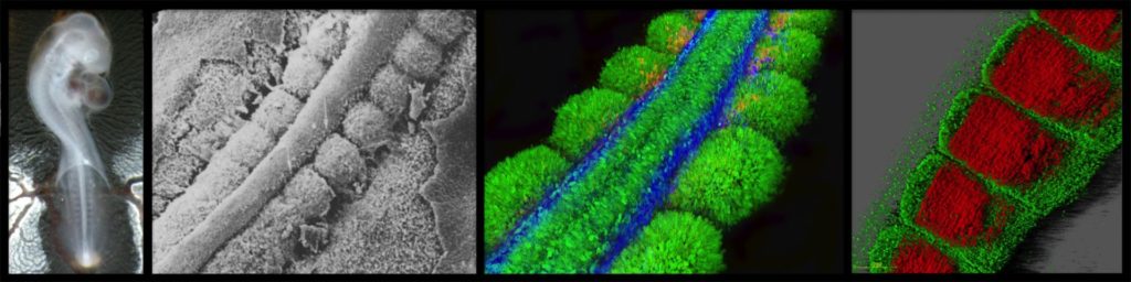
Embryon de poulet à 5,5 jours de développement, clarifié par la technique « 3DISCO » et observé au microscope à feuillet de lumière (Z1 Zeiss, CIQLE). Vert: crête neurale et système nerveux périphérique (anti-HNK1); Bleu: dermomyotome, progéniteurs musculaires et tube neural dorsal (anti-PAX7); Rouge: muscles différenciés (chaîne lourde anti-myosine). Marie-Julie Dejardin & Christophe Marcelle.
Ce film d’animation montre la morphogenèse et la croissance du myotome précoce (c’est-à-dire le muscle primitif) dans un embryon de poulet. Tous les muscles du corps et des membres dérivent de somites – des boules de cellules épithéliales qui se forment séquentiellement des deux côtés du tube neural au fur et à mesure du développement de l’embryon. On voit ici le compartiment dorsal des somites, appelé le dermomyotome, dont dérivent les muscles du tronc. Dans un premier temps, les cellules des lèvres médiale, postérieure, antérieure et enfin latérale du somite transloquent sous le dermomyotome, où elles s’allongent parallèlement à l’axe antéro-postérieur de l’embryon. Ces cellules post-mitotiques mononucléées et allongées sont appelées myocytes, et forment ensemble ce que nous appelons le myotome primaire. Dans un deuxième temps, la partie centrale du dermomyotome épithélial subit une transition épithélio-mensenchymateuse. En conséquence, une partie des cellules du dermomyotome migre vers l’ectoderme pour former plus tard le derme, tandis que d’autres cellules sont « parachutées » dans le myotome primaire. Contrairement aux myocytes qui ne se divisent pas, les cellules parachutées sont de véritables progéniteurs musculaires et peuvent se différencier ou s’autorenouveler. Grâce à ce processus, les muscles peuvent se développer pendant la vie embryonnaire et fœtale. Les cellules souches musculaires de l’adulte (appelées cellules satellites) proviennent de la même population de progéniteurs identifiée ici. Il est important de réaliser que le même processus morphogénétique a lieu chez la souris, et donc vraisemblablement chez l’homme. Ce film a été créé en 2005 par Jérôme Gros avec le logiciel gratuit open source 3D Blender. Publications associées : Gros, Scaal & Marcelle, Developmental Cell, 2004. Gros, Manceau, Thomé & Marcelle, Nature, 2005. Gros, Serralbo & Marcelle, Nature, 2009.
Transgenic quails reveal dynamic TCF/β-catenin signaling during avian embryonic development. Barzilai-Tutsch H, Morin V, Toulouse G, Chernyavskiy O, Firth S, Marcelle C, Serralbo O. Elife (2022) — Résumé
The Wnt/β-catenin signaling pathway is highly conserved throughout evolution, playing crucial roles in several developmental and pathological processes. Wnt ligands can act at a considerable distance from their sources and it is therefore necessary to examine not only the Wnt-producing but also the Wnt-receiving cells and tissues to fully appreciate the many functions of this pathway. To monitor Wnt activity, multiple tools have been designed which consist of multimerized Wnt signaling response elements (TCF/LEF binding sites) driving the expression of fluorescent reporter proteins (e.g. GFP, RFP) or of LacZ. The high stability of those reporters leads to a considerable accumulation in cells activating the pathway, thereby making them easily detectable. However, this makes them unsuitable to follow temporal changes of the pathway's activity during dynamic biological events. Even though fluorescent transcriptional reporters can be destabilized to shorten their half-lives, this dramatically reduces signal intensities, particularly when applied in vivo. To alleviate these issues, we developed two transgenic quail lines in which high copy number (12× or 16×) of the TCF/LEF binding sites drive the expression of destabilized GFP variants. Translational enhancer sequences derived from viral mRNAs were used to increase signal intensity and specificity. This resulted in transgenic lines efficient for the characterization of TCF/β-catenin transcriptional dynamic activities during embryogenesis, including using in vivo imaging. Our analyses demonstrate the use of this transcriptional reporter to unveil novel aspects of Wnt signaling, thus opening new routes of investigation into the role of this pathway during amniote embryonic development.
TGFβ signalling acts as a molecular brake of myoblast fusion. Melendez J, Sieiro D, Salgado D, Morin V, Dejardin MJ, Zhou C, Mullen AC, Marcelle C. Nature Communications (2021) — Résumé
Fusion of nascent myoblasts to pre-existing myofibres is critical for skeletal muscle growth and repair. The vast majority of molecules known to regulate myoblast fusion are necessary in this process. Here, we uncover, through high-throughput in vitro assays and in vivo studies in the chicken embryo, that TGFβ (SMAD2/3-dependent) signalling acts specifically and uniquely as a molecular brake on muscle fusion. While constitutive activation of the pathway arrests fusion, its inhibition leads to a striking over-fusion phenotype. This dynamic control of TGFβ signalling in the embryonic muscle relies on a receptor complementation mechanism, prompted by the merging of myoblasts with myofibres, each carrying one component of the heterodimer receptor complex. The competence of myofibres to fuse is likely restored through endocytic degradation of activated receptors. Altogether, this study shows that muscle fusion relies on TGFβ signalling to regulate its pace.
Transgenesis and web resources in quail. Serralbo O, Salgado D, Véron N, Cooper C, Dejardin MJ, Doran T, Gros J, Marcelle C. Elife (2020) — Résumé
Due to its amenability to manipulations, to live observation and its striking similarities to mammals, the chicken embryo has been one of the major animal models in biomedical research. Although it is technically possible to genome-edit the chicken, its long generation time (6 months to sexual maturity) makes it an impractical lab model and has prevented it widespread use in research. The Japanese quail (Coturnix coturnix japonica) is an attractive alternative, very similar to the chicken, but with the decisive asset of a much shorter generation time (1.5 months). In recent years, transgenic quail lines have been described. Most of them were generated using replication-deficient lentiviruses, a technique that presents diverse limitations. Here, we introduce a novel technology to perform transgenesis in quail, based on the in vivo transfection of plasmids in circulating Primordial Germ Cells (PGCs). This technique is simple, efficient and allows using the infinite variety of genome engineering approaches developed in other models. Furthermore, we present a website centralizing quail genomic and technological information to facilitate the design of genome-editing strategies, showcase the past and future transgenic quail lines and foster collaborative work within the avian community.
Cytoplasmic NOTCH and membrane derived β-catenin link fate choice to epithelial-mesenchymal transition during myogenesis. Sieiro D, Rios AC, Hirst CE, Marcelle C. Elife (2016) — Résumé
How cells in the embryo coordinate epithelial plasticity with cell fate decision in a fast changing cellular environment is largely unknown. In chick embryos, skeletal muscle formation is initiated by migrating Delta1-expressing neural crest cells that trigger NOTCH signaling and myogenesis in selected epithelial somite progenitor cells, which rapidly translocate into the nascent muscle to differentiate. Here, we uncovered at the heart of this response a signaling module encompassing NOTCH, GSK-3β, SNAI1 and β-catenin. Independent of its transcriptional function, NOTCH profoundly inhibits GSK-3β activity. As a result SNAI1 is stabilized, triggering an epithelial to mesenchymal transition. This allows the recruitment of β-catenin from the membrane, which acts as a transcriptional co-factor to activate myogenesis, independently of WNT ligand. Our results intimately associate the initiation of myogenesis to a change in cell adhesion and may reveal a general principle for coupling cell fate changes to EMT in many developmental and pathological processes.
A dynamic analysis of muscle fusion in the chick embryo. Sieiro-Mosti D, De La Celle M, Pele M, Marcelle C. Development (2014) — Résumé
Skeletal muscle development, growth and regeneration depend upon the ability of muscle cells to fuse into multinucleated fibers. Surprisingly little is known about the cellular events that underlie fusion during amniote development. Here, we have developed novel molecular tools to characterize muscle cell fusion during chick embryo development. We show that all cell populations arising from somites fuse, but each with unique characteristics. Fusion in the trunk is slow and independent of fiber length. By contrast, the addition of nuclei in limb muscles is three times more rapid than in trunk and is tightly associated with fiber growth. A complex interaction takes place in the trunk, where primary myotome cells from the medial somite border rarely fuse to one another, but readily do so with anterior and posterior border cells. Conversely, resident muscle progenitors actively fuse with one another, but poorly with the primary myotome. In summary, this study unveils an unexpected variety of fusion behaviors in distinct embryonic domains that is likely to reflect a tight molecular control of muscle fusion in vertebrates.
Migrating cells mediate long-range WNT signaling. Serralbo O, Marcelle C. Development (2014) — Résumé
In amniotes, it is widely accepted that WNTs secreted by the dorsal neural tube form a concentration gradient that regulates early somite patterning and myotome organization. Here we demonstrate in the chicken embryo that WNT protein is not secreted to act at a distance, but rather loaded onto migrating neural crest cells that deliver it to somites. Inhibiting neural crest migration or ablating their population has a profound impact on the WNT response in somites. Furthermore, we show that a central player in the efficient delivery of WNT to somites is the heparan sulfate proteoglycan GPC4, expressed by neural crest. Together, our data describe a novel mode of signaling whereby WNT proteins hitch a ride on migratory neural crest cells to pattern the somites at a distance from its source.
Neural crest regulates myogenesis through the transient activation of Notch. Rios AC, Serralbo O, Salgado D, Marcelle C. Nature (2011) — Résumé
How dynamic signalling and extensive tissue rearrangements interact to generate complex patterns and shapes during embryogenesis is poorly understood. Here we characterize the signalling events taking place during early morphogenesis of chick skeletal muscles. We show that muscle progenitors present in somites require the transient activation of NOTCH signalling to undergo terminal differentiation. The NOTCH ligand Delta1 is expressed in a mosaic pattern in neural crest cells that migrate past the somites. Gain and loss of Delta1 function in neural crest modifies NOTCH signalling in somites, which results in delayed or premature myogenesis. Our results indicate that the neural crest regulates early muscle formation by a unique mechanism that relies on the migration of Delta1-expressing neural crest cells to trigger the transient activation of NOTCH signalling in selected muscle progenitors. This dynamic signalling guarantees a balanced and progressive differentiation of the muscle progenitor pool.
Wnt11 acts as a directional cue to organize the elongation of early muscle fibers. Gros J, Serralbo O, Marcelle C. Nature (2009) — Résumé
The early vertebrate skeletal muscle is a well-organized tissue in which the primitive muscle fibres, the myocytes, are all parallel and aligned along the antero-posterior axis of the embryo. How myofibres acquire their orientation during development is unknown. Here we show that during early chick myogenesis WNT11 has an essential role in the oriented elongation of the myocytes. We find that the neural tube, known to drive WNT11 expression in the medial border of somites, is necessary and sufficient to orient myocyte elongation. We then show that the specific inhibition of WNT11 function in somites leads to the disorganization of myocytes. We establish that WNT11 mediates this effect through the evolutionary conserved planar cell polarity (PCP) pathway, downstream of the WNT/beta-catenin-dependent pathway, required to initiate the myogenic program of myocytes and WNT11 expression. Finally, we demonstrate that a localized ectopic source of WNT11 can markedly change the orientation of myocytes, indicating that WNT11 acts as a directional cue in this process. All together, these data show that the sequential action of the WNT/PCP and the WNT/beta-catenin pathways is necessary for the formation of fully functional embryonic muscle fibres. This study also provides evidence that WNTs can act as instructive cues to regulate the PCP pathway in vertebrates.
Myostatin promotes the terminal differentiation of embryonic muscle progenitors. Manceau M, Savage K, Gros J, Thome V, McPherron A, Paterson B, Marcelle C. Genes Dev (2009) — Résumé
Myostatin, a TGF-beta family member, is an important regulator of adult muscle size. While extensively studied in vitro, the mechanisms by which this molecule mediates its effect in vivo are poorly understood. We addressed this question using chick and mouse embryos. We show that while myostatin overexpression in chick leads to an exhaustion of the muscle progenitor population that ultimately results in muscle hypotrophy, myostatin loss of function in chick and mouse provokes an expansion of this population. Our data demonstrate that myostatin acts in vivo to regulate the balance between proliferation and differentiation of embryonic muscle progenitors by promoting their terminal differentiation through the activation of p21 and MyoD. Previous studies have suggested that myostatin imposes quiescence on muscle progenitors. Our data suggest that myostatin's effect on muscle progenitors is more complex than previously realized and is likely to be context-dependent. We propose a novel model for myostatin mode of action in vivo, in which myostatin affects the balance between proliferation and differentiation of embryonic muscle progenitors by enhancing their differentiation.
A Common Somitic Origin for Embryonic Muscle Progenitors and Satellite cells. Gros J, Manceau M, Thome V, Marcelle C. Nature (2005) — Résumé
In the embryo and in the adult, skeletal muscle growth is dependent on the proliferation and the differentiation of muscle progenitors present within muscle masses. Despite the importance of these progenitors, their embryonic origin is unclear. Here we use electroporation of green fluorescent protein in chick somites, video confocal microscopy analysis of cell movements, and quail-chick grafting experiments to show that the dorsal compartment of the somite, the dermomyotome, is the origin of a population of muscle progenitors that contribute to the growth of trunk muscles during embryonic and fetal life. Furthermore, long-term lineage analyses indicate that satellite cells, which are known progenitors of adult skeletal muscles, derive from the same dermomyotome cell population. We conclude that embryonic muscle progenitors and satellite cells share a common origin that can be traced back to the dermomyotome.
A two step mechanism for myotome formation in chick. Gros J, Scaal M, Marcelle C. Dev Cell (2004) — Résumé
The study of the morphogenetic cell movements underlying myotome formation in the chick embryo has led to the emergence of highly controversial models. Here we report a real-time cell lineage analysis of myotome development using electroporation of a GFP reporter in newly formed chick somites. Confocal analysis of cell movements demonstrates that myotome formation involves two sequential steps. In a first phase, incremental myotome growth results from a contribution of myocytes derived solely from the medial border of the dermomyotome. In a second phase, myocytes are produced from all four borders of the dermomyotome. The relative distribution of myocytes demonstrates that the medial and the lateral borders of the somite generate exclusively epaxial and hypaxial muscles. This analysis also identified five myotomal regions, characterized by the origin of the myocytes that constitute them. Together, our results provide a comprehensive model describing the morphogenesis of the early myotome in higher vertebrates.
Financements et soutien
ANR (2024-2028) : Muscle morphogenesis in the embryo (Myoptah), in collaboration with Pascal Maire (Institut Cochin).
AFM Téléthon (2025) : Epaxial Muscle Patterning
AFM-MyoNeurALP2 program (2022-2026) : Muscle fusion
Association Monégasque contre les Myopathies (2022-2025)
ANR (2022-2026) : Fusion of T-cells to muscle to alleviate dystrophies. ANR Partners : Frederic Relaix (Institut Mondor de recherche biomedical) , Luis Garcia (UVSQ-Université Paris-Saclay).